


Excellent cutting
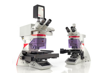
laser micro cutting Leica LMD6 & Lmd7
different from the traditional laser micro cutting system, Leica laser micro cutting system does not need to move the sample, but through moving the laser and gravity collection, to avoid sample pollution to a large extent, and provide you with an ideal cutting tissue sample for real-time analysis
laser microdissection (LMD), also known as laser capture microdissection or LCM, facilitates the user to separate specific individual cells or entire tissue areas. Leica laser microdissection system adopts unique laser design and easy-to-use dynamic software. From the whole tissue area to a single cell, users can easily separate the target area (ROI)
laser microdissection is commonly used in genomics (DNA), transcriptomics (mRNA, miRNA), proteomics, metabolomics, and even next generation sequencing (NGS). Neuroscience, cancer research, plant analysis, forensic medicine or climate researchers all use this microdissection technology to conduct discipline research. In addition, laser microdissection is also an ideal tool for living cell culture (LCC), which can be used for cloning, re culture, manipulation or downstream analysis

Leica laser capture micro cutting provides objective lens with different magnification
excellent performance
since the early 19th century, the development and manufacturing of optical components has been an important part of our core competitiveness. Please rely on the excellent performance of Leica smartcut series laser micro cutting special objective lens, which can help you achieve excellent cutting effect
selection range: 10 kinds of dry objective, from 5x to 150x
excellent imaging performance, when necessary, the unique 150x smartcut objective can be used to observe the sample details at high magnification and high resolution
larger field of view can be obtained by using low magnification objective, By cutting large samples intact
the laser transmittance of the objective lens can reach 350 nm, which can complete the laser micro cutting of tissues, bones, teeth, brain, plants, chromosomes and living cells
the same principle, two sets of systems
working tools: Leica lmd6 and Leica lmd7, the main difference between the two products is laser. Leica lmd6 is an ideal tool for standard application of dissecting soft tissues such as brain, liver or kidney. Leica lmd7 can ideally cut any type, size or shape of tissue. Leica lmd7 offers greater flexibility, higher laser power and more laser controls than smaller systems
two systems for your exploration:
Leica lmd6 can achieve excellent results in standard tissue cutting
Leica lmd7 can meet high expectations and flexible use
uniform lighting
light is crucial when defining the cutting area. The Leica lmd6 and Leica lmd7 laser micro cutting systems can be illuminated by traditional halogen lamps or LED
LED Benefits of lighting:
under sufficient lighting, you can see the natural color of the sample - because LED lighting can illuminate the sample evenly and has a constant color temperature
LED lighting can save 90% of energy and has up to 25, 000 hours of service life – instrument downtime caused by bulb replacement has long been a thing of the past
If halogen lighting for transmitted light is still your preferred choice, our two systems can also be equipped. We can provide internal constant color temperature control (CCIC) to avoid any image changes caused by traditional lighting technology, even if the system is used in applications unrelated to laser micro cutting
overview of advanced laser technology
| Leica LMD6 | Leica Lmd7 | |
| wavelength | 355 nm | 349 nm |
| pulse frequency | 80 Hz | 10-5000 Hz |
| pulse length | & lt; 4 ns | < 4 ns |
| maximum pulse energy | 70 µ J | 120 µ J |
Leica LMD7 Humanized design, convenient and easy to use:
it integrates the high energy and high and adjustable repetition rate of each pulse into a system
you can completely control the repetition rate, And adjust the laser speed according to specific samples
you can use the high energy of each pulse for thick and hard samples
to enjoy the convenience of high speed and high repetition rate in narrow cutting
you can control all laser parameters including laser aperture to achieve the appropriate cutting line
Save consumables
because Leica laser microdissection system only uses gravity to collect and Excise tissues, you can use all the common molecular biological reaction devices, such as the 0.2 or 0.5 ml cap in your laboratory, to save consumables. In addition, the collection device can be either dry or a reaction buffer or culture medium for LMD application
in the "mobile cutting" mode, the film slide was used to achieve the ideal result: the tissue was cut directly in the field. This method is called laser microdissection, which is an effective and time-saving method to obtain ideal image quality
use "draw scan" mode to cut on ordinary glass slide, cover slide or director slide: this method is called laser ablation or point scan cutting, and can be used for film free collection
The Leica laser microdissection system adopts a vertical optical imaging system to provide you with a high-resolution field of vision and ensure the accurate differentiation and cutting of living tissues, cells and even subcellular structures
intuitive and easy-to-use software makes laser micro cutting more convenient
Leica laser micro system software is easy to use and powerful, easy to select, cut and visually cut tissues, providing you with intuitive cutting results
a variety of slide solutions to meet a variety of needs
if you are engaged in proteomics or metabolomics, perhaps the plasticizer or softener of film slide may interfere with your analysis. Therefore, Leica micro system provides a variety of slide options for laser micro cutting
any kind of film slide can be used for genomics and transcriptomics
Pet slide can be used for specific applications in proteomics and metabolomics, and pet almost does not contain softener
the director slide can be selected for proteomics and metabolomics, so that it can be used completely without thin film
can overview the sample for better positioning
use mouse or touch screen to guide laser beam
control laser and microscope
record time-lapse image
such as data base, automatic cell recognition (AVC Pattern recognition) and other additional software packages and more features are available at any time. Sales representatives understand our comprehensive service
ultimate goal: save time and effort
you can cut the living cells in the culture bacteria to re culture, clone or analyze individual cells Colonies or cell groups
you can even connect the climate chamber to the laser microdissection system
you can use pen film or multi plate ibidi slide to cultivate cells in the culture dish
you can collect the excised tissue of living cell culture bacteria into the culture dish (with or without pen film, ibidi slide, or 8-stripe tube), Or collect it to PCR cap and other collection equipment for analysis
